Understanding Adrenal CT: A Comprehensive Guide to Imaging Techniques
The adrenal glands, small but vital organs located atop each kidney, play a crucial role in the body’s endocrine system. They are responsible for producing a variety of hormones, including adrenaline, cortisol, and aldosterone, which regulate numerous physiological processes such as metabolism, immune response, and stress management. Due to their importance, any abnormalities or diseases affecting the adrenal glands can have significant implications for overall health.
Imaging techniques, particularly computed tomography (CT), have become essential tools in diagnosing and managing conditions related to the adrenal glands. These advanced imaging methods allow healthcare professionals to visualize the structure and function of the adrenal glands, helping to identify tumors, hyperplasia, and other disorders. Understanding the various CT techniques and their applications can empower patients and healthcare providers alike, ensuring more informed decisions regarding diagnosis and treatment.
This comprehensive guide delves into the intricacies of adrenal CT imaging, providing insights into the techniques, benefits, and considerations involved. By exploring this topic, readers can gain a better understanding of how these imaging techniques contribute to adrenal health and disease management.
What is Adrenal CT Imaging?
Adrenal CT imaging is a specialized radiological technique used to visualize the adrenal glands and surrounding structures. It employs advanced X-ray technology to create detailed cross-sectional images of the body’s internal organs. The primary purpose of adrenal CT is to detect abnormalities within the adrenal glands, such as tumors, cysts, or enlargement due to hyperplasia.
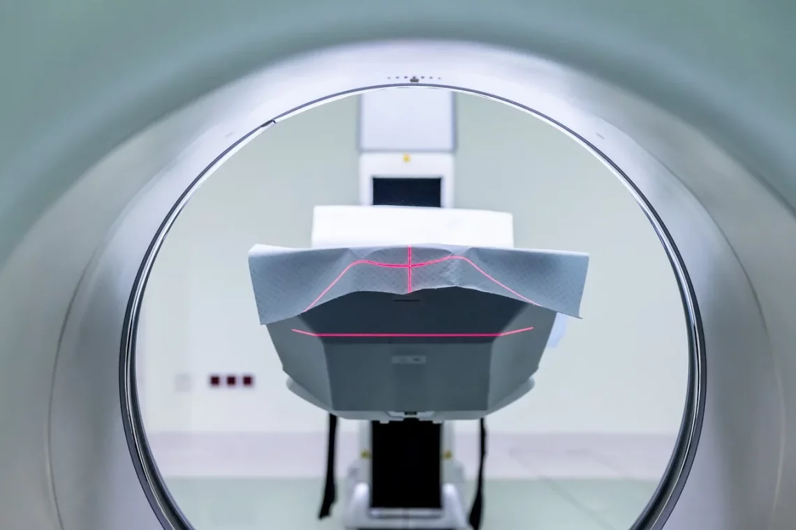
CT scans work by using ionizing radiation to produce images. The patient lies on a table that moves through a large, donut-shaped machine, which takes a series of X-rays from different angles. These images are then processed by a computer to generate cross-sectional views of the adrenal glands. Because of their high-resolution capabilities, CT scans are particularly useful for identifying small lesions that might be missed by other imaging modalities.
There are several types of CT scans utilized for adrenal imaging, including non-contrast, contrast-enhanced, and adrenal protocol CT. Non-contrast CT scans are typically the first step in evaluating adrenal abnormalities, as they provide a baseline image. However, contrast-enhanced CT scans offer enhanced visualization of the adrenal glands, allowing for better assessment of blood flow and tissue characteristics. The adrenal protocol CT, often performed after the administration of intravenous contrast, is specifically tailored to evaluate the adrenal glands and may include additional imaging sequences to improve diagnostic accuracy.
In addition to their diagnostic capabilities, adrenal CT scans are instrumental in guiding treatment decisions. For instance, if a tumor is detected, the imaging results can help determine the best course of action, whether that be surgical intervention, monitoring, or medical management.
The Role of Contrast in Adrenal CT
Contrast agents are substances used in various imaging studies to improve the visibility of specific organs or tissues. In adrenal CT imaging, contrast plays a vital role in enhancing the quality of the images obtained. By increasing the difference in density between the adrenal glands and surrounding tissues, contrast agents help radiologists identify abnormalities more effectively.
There are two primary types of contrast agents used in CT imaging: iodinated contrast media and negative contrast agents. Iodinated contrast media, which contain iodine, are the most commonly used for adrenal CT scans. These agents are administered intravenously and circulate through the bloodstream, allowing for better visualization of vascular structures and enhancing the delineation of the adrenal glands.
The timing of contrast administration is crucial for optimal imaging results. Typically, the contrast is injected shortly before the scan begins, allowing it to distribute within the bloodstream. Radiologists often use specific timing protocols to capture images at various phases after contrast injection, known as arterial, venous, and delayed phases. Each phase provides different information about the vascularity and characteristics of the adrenal glands, aiding in the differentiation between benign and malignant lesions.
While the use of contrast agents significantly enhances the diagnostic capabilities of adrenal CT, it’s essential to consider potential risks. Some patients may experience allergic reactions to iodinated contrast media, while others may have kidney function concerns that could be exacerbated by contrast administration. Therefore, healthcare providers must assess each patient’s medical history and current health status before proceeding with contrast-enhanced imaging.
Indications for Adrenal CT Scans
Adrenal CT scans are indicated for various clinical scenarios, primarily focused on the evaluation of adrenal abnormalities. Patients may be referred for adrenal CT imaging based on symptoms, physical examination findings, or laboratory results that suggest adrenal dysfunction.
One of the most common indications for adrenal CT is the evaluation of adrenal masses. When imaging studies or laboratory tests raise concerns about the presence of a tumor, a CT scan can help characterize the mass, assess its size, and determine its relationship to surrounding structures. This information is crucial for establishing a diagnosis and formulating a treatment plan.
In addition to tumor detection, adrenal CT is also used to evaluate conditions such as Cushing’s syndrome, which results from excess cortisol production, and Conn’s syndrome, characterized by excessive aldosterone secretion. In these cases, imaging studies help identify potential adrenal adenomas or hyperplasia that may be responsible for hormone overproduction.
Moreover, adrenal CT scans are frequently employed in the staging of adrenal cancer, particularly in cases of metastatic disease. Understanding the extent of cancer spread is vital for developing an effective treatment strategy, and CT imaging provides essential information about lymph node involvement and distant metastases.
Lastly, adrenal CT can aid in postoperative assessment following adrenal surgery. After surgical intervention, imaging studies are often performed to monitor for complications, such as bleeding or infection, and to evaluate the success of the procedure in removing the targeted tissue.
Future Trends in Adrenal Imaging
As technology continues to advance, the field of adrenal imaging is evolving with new techniques and modalities that enhance diagnostic accuracy and patient safety. One of the most promising developments is the integration of artificial intelligence (AI) into imaging analysis. AI algorithms can aid radiologists in interpreting complex imaging studies, improving the detection of subtle lesions and reducing the risk of human error.
Additionally, advancements in imaging technology, such as multi-detector CT and high-resolution imaging, offer improved visualization of the adrenal glands. These innovations allow for faster scans with greater detail, enabling better diagnosis and treatment planning.
Another trend is the growing interest in functional imaging, which assesses not only the anatomical structure of the adrenal glands but also their metabolic activity. Techniques such as positron emission tomography (PET) imaging, often combined with CT, provide insights into the functional status of adrenal lesions, helping to differentiate between benign and malignant processes.
Moreover, the development of targeted therapies for adrenal tumors is changing the landscape of treatment options. As more is understood about the genetic and molecular characteristics of adrenal tumors, imaging studies will play a crucial role in guiding personalized treatment strategies.
In conclusion, adrenal CT imaging is a vital tool in the assessment of adrenal health, offering detailed insights into the structure and function of these critical glands. With ongoing advancements in technology and imaging techniques, the future of adrenal imaging looks promising, ultimately leading to better patient outcomes.
**Disclaimer:** This article is intended for informational purposes only and does not constitute medical advice. For any health concerns or medical conditions, please consult a qualified healthcare professional.


